
You find a lump on your dog. It has been there for a few weeks. It seems to change in size. You are not sure whether to worry.
If that sounds familiar, you are not alone. Mast cell tumours are the most commonly diagnosed cutaneous (skin) cancer in dogs and they are also one of the most misunderstood. A mast cell tumour on a dog can look completely benign, shrink and swell unpredictably, and yet in some cases behave with considerable aggression.
The reassuring truth is that many mast cell tumours in dogs are low-grade and surgically curable. The critical truth is that you cannot tell which kind you are dealing with just by looking. Only a veterinary diagnosis can determine that and the earlier it happens, the more treatment options are available.
This article explains everything you need to know: what mast cell tumours are, how they are graded and staged, what treatment involves, and what the prognosis looks like across different grades and presentations.
Why Mast Cell Tumours Are One of the Most Common Skin Cancers in Dogs
Mast cells are a normal and essential part of the immune system. They are found throughout the body but are particularly concentrated in the skin, gastrointestinal tract, and respiratory tract tissues that interact regularly with the outside world.
Their job is to respond to injury, allergens, and parasites. They do this by releasing powerful chemical mediators most notably histamine, heparin, and proteases stored in granules inside the cell.
A mast cell tumour (MCT), also called a mastocytoma, forms when mast cells undergo abnormal, uncontrolled proliferation. Because the skin is so densely populated with mast cells, it is the most common site for these tumours to develop.
Mast cell tumours account for approximately 16–21% of all cutaneous tumours diagnosed in dogs. They occur across all breeds and ages, but certain breeds particularly those with a history of selective breeding carry a significantly higher genetic risk.
Terminology Note: You may see this condition written as mast cell tumour, mast cell tumor, mastocytoma, or occasionally as a mast cell tumor mastocytoma in dogs these all refer to the same condition. The British spelling uses ‘tumour’; the American spelling uses ‘tumor’. Both are correct.
Suggested Read: Early Signs of Cancer in Dogs You Should Watch Out For
How Mast Cell Tumours Develop in Dogs
The exact trigger for malignant transformation of mast cells is not fully understood, but research has identified several contributing factors.
Genetic and Molecular Factors
- KIT gene mutations: A mutation in the c-KIT proto-oncogene (which encodes the KIT receptor tyrosine kinase) is identified in approximately 15–40% of canine mast cell tumours. This mutation causes constitutive (unregulated) activation of KIT signalling, driving abnormal cell proliferation and survival.
- Breed predisposition: Certain breeds have heritable predispositions, suggesting that genetic factors play a significant role independent of environmental triggers.
- Chromosomal abnormalities: Alterations in tumour suppressor genes and oncogenes have been documented in high-grade tumours.
Breed Predisposition for Mast Cell Tumours in Dogs
The following breeds are recognised as being at significantly elevated risk:
- Boxers among the highest risk of any breed, though they tend to develop lower-grade tumours
- Bulldogs (English and French)
- Boston Terriers
- Labrador Retrievers
- Golden Retrievers
- Pugs
- Cocker Spaniels
- Shar-Peis tend to develop mast cell tumours at a younger age and sometimes with more aggressive behaviour
- Weimaraners
Mixed breed dogs are not immune, mast cell tumours occur in crossbreeds as well, though typically at lower rates than the predisposed breeds listed above.
What a Mast Cell Tumour Looks Like on a Dog
This is where mast cell tumours earn their reputation as the ‘great pretenders’ of canine oncology. They have no consistent appearance. A mast cell tumour on a dog can look like almost anything.
Common Presentations
- A raised, firm, hairless skin nodule the most stereotypical appearance
- A soft, fluctuant lump that feels almost like a lipoma (fatty lump)
- A flat, erythematous (reddened) skin lesion with poorly defined edges
- A pedunculated (stalked) skin tag-like growth
- An ulcerated or crusted lesion that looks like a wound that will not heal
- A wart-like growth, especially in short-coated breeds
The Darier Sign of Mast Cell Tumours
One characteristic feature worth knowing is the Darier sign. When a mast cell tumour is manipulated, scratched, or even just stroked, it may suddenly become red, swollen, and inflamed sometimes within minutes. This happens because physical stimulation degranulates the tumour cells, releasing histamine locally. The lump appears to dramatically ‘flare up’ when touched.
If a lump on your dog repeatedly becomes red and swollen, then reduces again over hours especially when scratched or irritated this pattern should be discussed with your vet promptly.
Variable Behaviour in Size
Many owners report that the lump seems to ‘come and go’ or change in size from day to day. This is not your imagination. Mast cell tumours can genuinely fluctuate in size due to intermittent degranulation and local inflammatory responses. A lump that changes in size is not reassuring it is a reason to investigate further, not a reason to wait.
Common Locations of Mast Cell Tumours in Dogs
- Trunk and limbs the most common sites
- Inguinal region (groin) and perineum
- Head and neck
- Muzzle and lips
- Digital (toe) mast cell tumours tend to have a more aggressive behaviour
- Subcutaneous (beneath the skin) these can be mistaken for benign fatty lumps
Key Point: There is no ‘typical’ appearance for a mast cell tumour on a dog. Any lump that changes size, becomes intermittently inflamed, or simply does not go away after four weeks should be assessed by a vet. Do not wait and see.
Signs and Symptoms of Mast Cell Tumour in Dogs
The visible lump is usually the first and in low-grade cases, the only sign. However, when mast cell tumours are more advanced, locally infiltrative, or when they degranulate systemically, a broader range of symptoms can develop.
Local Signs
- A skin mass or nodule, present for weeks to months
- Intermittent redness, swelling, or itching at the tumour site
- Ulceration or bleeding from the surface of the lump
- Hair loss over the tumour
- Regional lymph node enlargement a swollen lymph node near the tumour site
Also Read: Histiocytomas in Dogs: Symptoms, Treatment, and More
Systemic Signs When Degranulation Becomes Systemic
In high-grade, widely metastatic, or heavily manipulated tumours, the mass release of histamine and other mediators can cause systemic effects. These are serious and require immediate veterinary attention.
- Gastrointestinal ulceration: Histamine stimulates gastric acid secretion, which can cause gastric and duodenal ulcers. This is one of the most clinically significant complications of mast cell tumour disease.
- Vomiting and diarrhoea: Often caused by GI ulceration or direct mast cell infiltration of the GI tract.
- Loss of appetite and weight loss: Associated with GI disease, nausea, or systemic disease burden.
- Anaphylactic reaction: Massive, sudden degranulation triggered by rough manipulation of the tumour, surgery without pre-medication, or spontaneous rupture can cause acute systemic hypotension, facial swelling, vomiting, and cardiovascular collapse. This is rare but a genuine risk.
- Lethargy and generalised weakness: In dogs with widespread metastatic disease.
How Vets Diagnose Mast Cell Tumours in Dogs
An accurate diagnosis is the essential first step before any treatment decision. The appearance of the lump alone is never sufficient to confirm a mast cell tumour.
Fine Needle Aspirate (FNA) Cytology
This is the most commonly used initial diagnostic test. A fine needle is inserted into the lump and cells are aspirated onto a microscope slide. The slide is stained and examined by a veterinary pathologist.
- Mast cells have a characteristic appearance: round cells with dark purple granules in the cytoplasm
- FNA is quick, minimally invasive, and can often be performed without sedation
- It can confirm that a lump is a mast cell tumour with high reliability
Limitation: FNA can diagnose the tumour type but cannot determine the histological grade. For grade, a proper tissue biopsy is needed.
Histopathology Tissue Biopsy
A tissue biopsy either an incisional (partial) or excisional (full removal) biopsy, provides a sample large enough for histological evaluation. This is necessary to:
- Determine the tumour grade (see next section)
- Assess whether surgical margins are clean (whether all the cancer was removed)
- Perform additional molecular tests such as KIT mutation analysis
Staging Diagnostics of MCT: Has It Spread?
- Regional lymph node aspiration or biopsy enlarged lymph nodes near the tumour should always be assessed
- Abdominal ultrasound to evaluate the liver, spleen, and abdominal lymph nodes for evidence of spread
- Chest radiographs to assess for pulmonary metastasis (less common with MCT but performed in high-grade cases)
- Buffy coat smear a blood test that looks for circulating mast cells, seen in advanced systemic mastocytosis
- Bone marrow aspirate in cases with suspected systemic spread
Understanding Mast Cell Tumour Grading in Dogs
Grading is the cornerstone of mast cell tumour management. It tells the oncologist and surgeon how aggressive the tumour is likely to behave and therefore what level of treatment is needed.
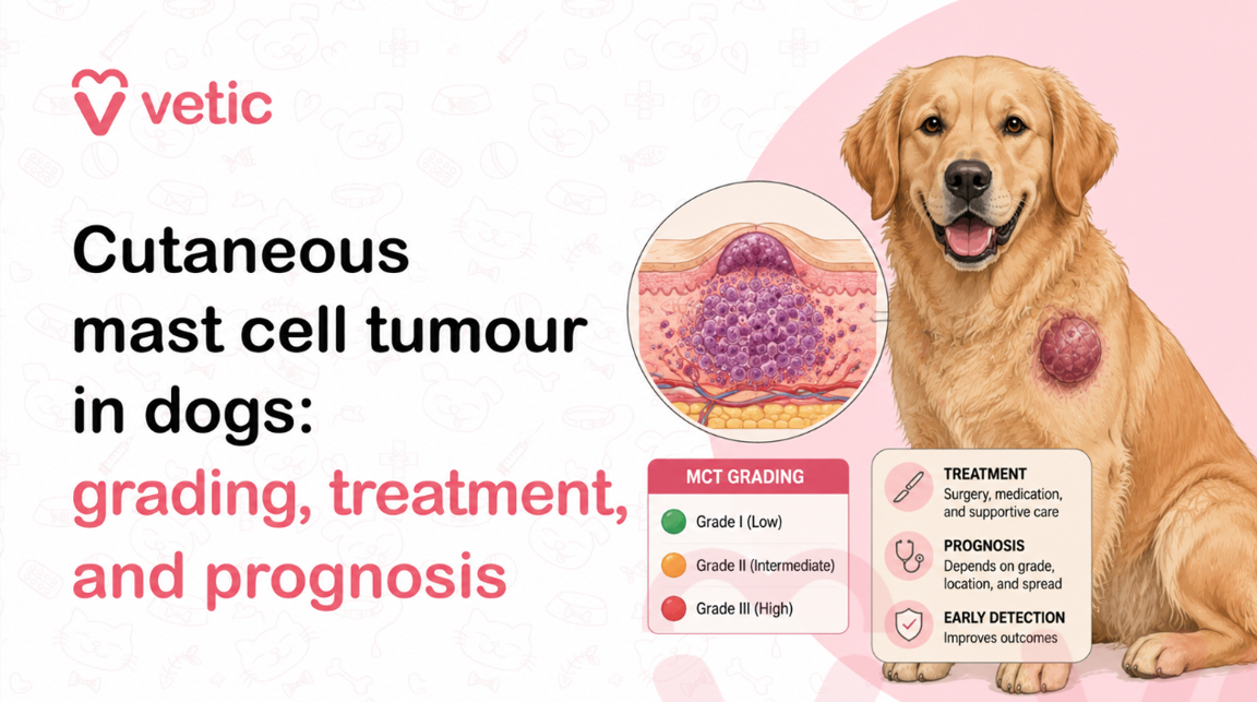
Two grading systems are currently used in veterinary oncology:
- The Patnaik system (3-tier): Grade I (benign), Grade II (intermediate), Grade III (malignant). Widely used for decades but with documented variability in inter-pathologist agreement, particularly for Grade II tumours.
- The Kiupel system (2-tier): Low-grade and High-grade. Developed more recently to improve consistency and provide clearer prognostic information. Now the preferred system in many specialist oncology practices.
Low-Grade vs High-Grade Mast Cell Tumours
| Feature | Low-Grade MCT | High-Grade MCT |
| Mitotic index | Low (fewer dividing cells) | High (many dividing cells) |
| Cell appearance | Well-differentiated, uniform | Poorly differentiated, pleomorphic |
| Behaviour | Locally invasive; slow to spread | Rapidly invasive; high metastatic potential |
| Proportion of MCTs | Approximately 70–75% of all MCTs | Approximately 25–30% of all MCTs |
| Surgical cure rate | High with wide clean margins | Lower; recurrence and spread are common |
| Median survival (post-surgery alone) | Greater than 2 years in most studies | 4–6 months without adjuvant treatment |
Why Tumour Grade Affects Treatment Decisions
Grade is not simply a label, it is the primary driver of every clinical decision that follows.
- Surgical margins: Low-grade tumours can often be managed with 2cm lateral margins. High-grade tumours require wider margins, and even then, adjuvant treatment is typically recommended.
- Need for adjuvant therapy: Low-grade tumours that are completely excised with clean margins often require no further treatment. High-grade tumours almost always warrant chemotherapy or targeted therapy.
- Monitoring intensity: High-grade disease requires more frequent follow-up and surveillance imaging.
- Owner counselling: Grade determines the realistic discussion about long-term outcomes including recurrence risk, quality of life, and when palliative management may become appropriate.
Important: A pathology report saying ‘Grade II’ under the Patnaik system can refer to tumours with widely different behaviours. If your dog’s report uses the Patnaik system, ask your vet or oncologist whether further risk stratification is possible, or whether Kiupel re-grading is an option.
Staging Mast Cell Tumours: Has the Cancer Spread?
Staging answers a different question from grading. Grading asks: how aggressive is this tumour at a cellular level? Staging asks: where has the disease gone?
The WHO staging system for canine mast cell tumours is as follows:
| Stage 0 | Single tumour incompletely excised, confirmed by histopathology, with no lymph node involvement. |
| Stage I | Single tumour confined to the dermis, with no lymph node involvement. |
| Stage II | Single tumour in the dermis with regional lymph node involvement. |
| Stage III | Multiple dermal tumours, or a single large, deeply infiltrating tumour, with or without lymph node involvement. |
| Stage IV | Any tumour with distant metastasis to the liver, spleen, bone marrow, blood, or other distant organs. |
Stage is closely tied to prognosis. Stage I and Stage II disease with complete surgical excision carries a significantly better long-term outlook than Stage III or Stage IV, where systemic spread has occurred.
It is worth noting that staging in practice is not always straightforward. Lymph node cytology can be difficult to interpret reactive lymph nodes can look abnormal without containing tumour cells, and early micrometastatic spread can be missed. Specialist interpretation is always preferable for borderline results.
Treatment Options for Mast Cell Tumour in Dogs
Treatment for a mast cell tumour in a dog is not a one-size-fits-all decision. It is shaped by tumour grade, stage, location, surgical accessibility, the dog’s overall health, and critically the owner’s goals and resources. The main treatment modalities are:
Surgery
Surgery is the treatment of first choice for localised, resectable mast cell tumours. The goal is complete excision with adequate margins meaning the surgeon removes not just the visible tumour, but a ring of apparently normal tissue around it to capture any microscopic tumour extension.
- Standard lateral margins: 2–3cm of grossly normal tissue around the tumour, where anatomically possible.
- Deep margin: One fascial plane deep to the tumour.
- Margin assessment: The excised tissue is submitted to a pathologist who evaluates the edges (margins) of the specimen. Clean margins (‘tumour-free margins’) indicate complete excision. Dirty or narrow margins indicate tumour cells may remain and further treatment is needed.
For tumours in surgically challenging locations the face, digits, prepuce, or perineum achieving wide margins may be difficult or impossible. In these cases, referral to a veterinary surgical specialist or oncologist is strongly advised.
Radiation Therapy
- Used as a primary treatment when surgery cannot achieve adequate margins
- Used adjunctively after surgery with incomplete margins in low- to intermediate-grade tumours
- Particularly effective for localised, non-metastatic disease where the tumour bed is accessible
- Requires specialist referral to a facility with a veterinary radiation oncology unit
- A course of radiation typically involves multiple fractions over three to four weeks
Medical Pre-treatment Before Surgery
Before any surgical procedure on a mast cell tumour, pre-medication is standard practice to reduce the risk of degranulation during surgery. This typically includes:
- H1 antihistamines block histamine H1 receptors
- H2 antihistamines block gastric histamine receptors to protect against ulceration
- These are continued post-operatively for a period determined by the surgeon
When Surgery Is Enough and When Additional Treatment Is Needed
This is one of the most important questions in mast cell tumour management and the answer comes from the pathology report.
| Low-grade + clean margins | Surgery is likely curative. Regular monitoring is recommended. No adjuvant therapy typically needed. |
| Low-grade + incomplete margins | Re-excision to achieve clean margins is the preferred option. Radiation therapy if re-excision is not possible. |
| High-grade + clean margins | Chemotherapy or targeted therapy strongly recommended even with clean margins, due to high metastatic risk. |
| High-grade + incomplete margins | Combination of re-excision or radiation AND systemic therapy. |
| Metastatic disease (any grade) | Surgery for local control (if appropriate) plus systemic therapy. Goals shift toward quality of life and disease control rather than cure. |
Vet Note: Do not rely on the appearance of margins at surgery what the surgeon sees is not sufficient. Always submit excised tissue for pathological margin assessment. A tumour that appears to have been cleanly removed can have microscopic tumour cells at the margins that only histopathology will reveal.
Role of Chemotherapy and Targeted Therapy in Dogs
For dogs with high-grade tumours, metastatic disease, or incomplete excision, systemic therapy extends beyond surgery.
Conventional Chemotherapy
- Vinblastine and prednisolone: The most widely used chemotherapy protocol for canine mast cell tumours. Vinblastine is given intravenously, typically weekly or biweekly, combined with oral prednisolone. This protocol is well-tolerated by most dogs.
- CCNU (lomustine): An oral alkylating agent used in dogs that have not responded to vinblastine, or as a rescue protocol in recurrent disease.
- Combination protocols: Various combinations of the above agents are used in specialist oncology settings, tailored to the individual patient.
Targeted Therapy Tyrosine Kinase Inhibitors (TKIs)
This is one of the most significant advances in canine oncology of the past two decades. Tyrosine kinase inhibitors (TKIs) are targeted drugs that specifically inhibit the abnormally activated KIT signalling pathway driving mast cell tumour growth.
- Toceranib phosphate (Palladia): The first cancer drug specifically approved for use in dogs. Approved for the treatment of recurrent or non-resectable Grade II/III mast cell tumours, particularly those with KIT mutations. Given orally.
- Masitinib (Masivet): Another TKI approved in some countries for canine MCT. Selectively targets the KIT receptor.
- Response rates: In dogs with KIT mutations, TKIs produce objective responses (measurable tumour reduction) in approximately 40–60% of cases. Disease stabilisation is observed in a further significant proportion.
TKIs are not without side effects gastrointestinal toxicity, protein-losing nephropathy, and neutropaenia can occur and require monitoring. But for dogs with KIT-mutant, high-grade, or non-resectable disease, they represent a meaningful treatment option.
Glucocorticoids (Steroids)
- Prednisolone alone can produce partial responses in some mast cell tumours particularly when combined with other agents
- Steroids reduce inflammation, stabilise mast cell membranes, and have direct anti-tumour activity in some cases
- Used as part of the vinblastine/prednisolone protocol and in palliative management
Prognosis: Life Expectancy with Mast Cell Tumour in Dogs
Prognosis for a dog with a mast cell tumour varies enormously from an excellent long-term outcome after a single surgical procedure, to a guarded prognosis requiring ongoing systemic therapy.
The most important determinants of prognosis are tumour grade and completeness of surgical excision.
| Scenario | Expected Outcome | Key Consideration |
| Low-grade, completely excised | Excellent cure is a realistic goal. Recurrence rates are low with clean margins. | Regular monitoring to catch any new or recurrent tumours early. |
| Low-grade, incompletely excised | Good with re-excision or radiation. Without further treatment, local recurrence is likely. | Do not leave incomplete excisions unaddressed. |
| High-grade, completely excised + adjuvant chemo | Guarded to fair. Median survival times of 12–18 months are reported in some studies with aggressive management. | KIT mutation status and metastatic spread significantly affect outcomes. |
| High-grade with regional lymph node involvement | Guarded. Survival is shortened compared to local disease alone. | Systemic therapy is essential. |
| Metastatic (Stage IV) | Poor. Goals shift to quality of life and disease control. Measured in months. | Palliative care and supportive management are appropriate discussions. |
Boxers deserve a specific mention: they are predisposed to mast cell tumours but tend to develop lower-grade disease. Multiple tumours are common in Boxers, but the individual tumour behaviour is typically less aggressive and therefore long-term outcomes are generally more favourable than in other breeds presenting with high-grade MCT.
Factors That Affect Survival in Dogs with Mast Cell Tumours
No two dogs and no two mast cell tumours are identical. Beyond grade and stage, several additional factors influence how a dog’s disease will behave over time.
- Tumour location: Mast cell tumours on the digits (toes), muzzle, preputial region, inguinal area, and perineum tend to behave more aggressively than those on the trunk, regardless of histological grade. Digit MCTs in particular carry a guarded prognosis.
- Mitotic index: Even within a grade, a high number of mitotic figures per high-power field is a powerful indicator of aggressive biological behaviour and shorter survival.
- Tumour size: Larger tumours are associated with a worse prognosis, partly because of the difficulty in achieving adequate surgical margins and partly because size correlates with disease burden.
- Rate of growth: A tumour that has appeared and grown rapidly over days to weeks is more concerning than one that has been stable for many months although even slow-growing tumours should not be left uninvestigated.
- Age and overall health: Younger, otherwise healthy dogs generally tolerate more intensive treatment and tend to have better responses to therapy.
- Time to diagnosis and treatment: Earlier diagnosis consistently correlates with better outcomes across nearly all stages and grades.
When to Worry About a Lump on Your Dog
Not every lump is a mast cell tumour, and not every lump requires emergency action. But the default stance should be: any new lump on your dog deserves veterinary assessment within a reasonable timeframe.
Seek veterinary attention promptly if a lump on your dog shows any of the following:
- Has been present for more than four weeks without change
- Is growing in size
- Changes size day to day growing and shrinking unpredictably
- Becomes intermittently red, swollen, or itchy
- Is ulcerated, bleeding, or weeping
- Is firm, poorly defined, or appears attached to deeper tissue
- Is located in a region associated with more aggressive MCT behaviour the digit, groin, muzzle, or perineum
Seek same-day veterinary attention if your dog with a known or suspected mast cell tumour develops:
- Acute vomiting, especially if bloody
- Profound lethargy or weakness
- Swelling of the face or throat
- Sudden, extreme worsening of a known mass following rough handling or trauma this may signal a degranulation event
Never assume a lump is benign: Without cytology or histopathology, there is no way to know whether a lump is a fatty cyst, a sebaceous adenoma, or a mast cell tumour. The cost of a fine needle aspirate is small. The cost of delaying diagnosis of a high-grade tumour can be significant.
Final Thoughts: Early Detection Can Improve Outcomes in Mast Cell Tumours
Mast cell tumours are not a diagnosis that should cause immediate despair but they are not a diagnosis that should be taken lightly either.
The range of outcomes across this disease is genuinely wide. Some dogs are surgically cured in a single procedure and never have a recurrence. Others face a longer road involving chemotherapy, targeted therapy, and careful monitoring. And for a smaller proportion, the goal shifts from cure to quality of life.
What consistently improves outcomes across grades, stages, breeds, and locations is early detection and early treatment. A low-grade mast cell tumour found and surgically excised with clean margins at two centimetres is a very different clinical problem from the same tumour found at five centimetres, with regional lymph node involvement, after months of observation.
The single most impactful thing you can do as a dog owner is to examine your dog regularly, to know their body, and to act on any new lump rather than adopting a wait-and-see approach. In veterinary oncology, waiting rarely helps and often harms.
Find it early. Investigate it promptly. Treat it appropriately. That is the principle that gives dogs with mast cell tumours the best possible chance.
FAQ: Mast Cell Tumour in Dogs
Are mast cell tumours in dogs always cancerous?
Mast cell tumours are classified as malignant neoplasms meaning they are technically cancer. However, the word ‘cancer’ encompasses a vast range of biological behaviours, and mast cell tumours reflect that range precisely. A low-grade mast cell tumour that is completely surgically excised may behave for all practical purposes like a cured condition, with an excellent long-term outlook. A high-grade tumour, by contrast, can be aggressive, metastatic, and life-limiting. The critical point is that you cannot determine which type you are dealing with from appearance alone; histopathological grading is essential.
How fast do mast cell tumours grow in dogs?
Growth rate varies considerably and is one of the clinical features that makes mast cell tumours so difficult to assess by observation alone. Low-grade tumours may be present for months or even years with minimal change giving a false sense of security. High-grade tumours can appear and grow significantly within days to weeks. Adding to the confusion, mast cell tumours can fluctuate in apparent size due to intermittent degranulation, making a rapidly growing tumour appear stable and then suddenly enlarged. Rate of growth alone is therefore not a reliable guide; cytological or histological assessment is the only reliable way to determine how a specific tumour is likely to behave.
Can mast cell tumours in dogs be cured?
Yes a proportion of mast cell tumours in dogs are genuinely and completely curable with surgery alone. Low-grade tumours that are completely excised with adequate surgical margins have low recurrence rates and, in many cases, are never a problem again. The challenge is that not all tumours can be completely excised (due to location or size), and high-grade disease carries a much higher risk of systemic spread that makes cure less reliable. Even with high-grade tumours, some dogs achieve extended periods of remission with multimodal treatment. Cure as a realistic goal versus disease control as a realistic goal is a discussion best held with a veterinary oncologist once grading and staging results are available.
What is the survival rate for dogs with mast cell tumours?
There is no single survival rate; the figures vary dramatically by grade, stage, and treatment. Dogs with low-grade tumours and complete surgical excision have a median survival exceeding two years in most studies, with many dogs living significantly longer. Dogs with high-grade tumours and no adjuvant treatment have a median survival of approximately four to six months. With chemotherapy and/or targeted therapy, this extends meaningfully median survival times of twelve to eighteen months are reported in dogs with high-grade disease receiving aggressive management. Metastatic Stage IV disease carries the most guarded outlook, with survival measured in months even with treatment. These numbers are population medians, not individual predictions individual dogs frequently exceed or fall short of median survival times.
Is surgery necessary for mast cell tumour removal?
Once a mast cell tumour has been diagnosed or is strongly suspected based on cytology surgical removal is generally recommended without unnecessary delay, particularly for tumours that are growing, changing, or in a surgically accessible location. However, ‘immediately’ needs to be balanced with ‘adequately planned’. Rushing into surgery without appropriate pre-surgical histological diagnosis, staging, and pre-medication planning can lead to incomplete excision, inadequate margins, or complications from intraoperative degranulation. The right approach is: confirm the diagnosis via FNA cytology, complete appropriate staging diagnostics for high-grade or concerning tumours, pre-medicate appropriately, and perform definitive surgery with wide margins by a surgeon experienced in oncological technique or refer to a specialist. Urgency matters, but planning matters equally.
References:
Berlato, D., Bulman-Fleming, J., Clifford, C. A., Garrett, L., Intile, J., Jones, P., Kamstock, D. A., Liptak, J. M., Pavuk, A., Powell, R., Rasotto, R. (2021). Value, limitations, and recommendations for grading of canine cutaneous mast cell tumors: A consensus of the Oncology-Pathology Working Group. Veterinary Pathology, 58(5), 858–863. https://doi.org/10.1177/03009858211009785
Blackwood, L., Murphy, S., Buracco, P., De Vos, J. P., De Fornel-Thibaud, P., Hirschberger, J., Kessler, M., Pastor, J., Ponce, F., Savary-Bataille, K., & Argyle, D. J. (2012). European consensus document on mast cell tumours in dogs and cats. Veterinary and Comparative Oncology, 10(3), e1–e29. https://doi.org/10.1111/j.1476-5829.2012.00341.x
Kiupel, M., Webster, J. D., Bailey, K. L., Best, S., DeLay, J., Detrisac, C. J., Fitzgerald, S. D., Gamble, D., Ginn, P. E., & Goldschmidt, M. H. (2011). Proposal of a 2-tier histologic grading system for canine cutaneous mast cell tumors to more accurately predict biological behavior. Veterinary Pathology, 48(1), 147–155. https://doi.org/10.1177/0300985810386469
London, C. A., Malpas, P. B., Wood-Follis, S. L., Boucher, J. F., Rusk, A. W., Rosenberg, M. P., Henry, C. J., Mitchener, K. L., Klein, M. K., Hintermeister, J. G., Bergman, P. J., Couto, G. C., Mauldin, G. N., & Michels, G. M. (2009). Multi-center, placebo-controlled, double-blind, randomized study of oral toceranib phosphate (SU11654), a receptor tyrosine kinase inhibitor, for the treatment of dogs with recurrent mast cell tumor following surgical excision. Clinical Cancer Research, 15(11), 3856–3865. https://doi.org/10.1158/1078-0432.CCR-08-1860

