
Finding a sudden lump on your dog’s skin can be alarming. One common cause, especially in young dogs, is a histiocytoma. These growths often look aggressive and appear quickly. But the good news is that most histiocytomas are benign and self-limiting. Still, not every lump is harmless, and knowing what to watch for is essential.
This blog explains what dog histiocytomas are, how they look, why they form, available treatment options, including pet surgery, home care tips, and when a similar-looking lump may be a more serious condition.
What Are Histiocytomas in Dogs?
A histiocytoma in dogs is a benign (non-cancerous) skin tumour that arises from Langerhans cells, a type of immune cell found in the skin. These cells normally help protect the body from infection. In some dogs, particularly young ones, these cells can grow rapidly and form a small skin mass.
Canine histiocytomas are:
- Typically benign
- Most common in dogs under 2 years old
- Often solitary (just one lump)
- Frequently self-resolving within weeks to months
They are considered part of a broader category of skin conditions involving histiocytic cells. Importantly, they are different from malignant histiocytic disorders, which are rare but serious.
What Does a Canine Histiocytoma Look Like?
Histiocytomas have a fairly classic appearance, though they can still be mistaken for other skin tumours. Typical features include:
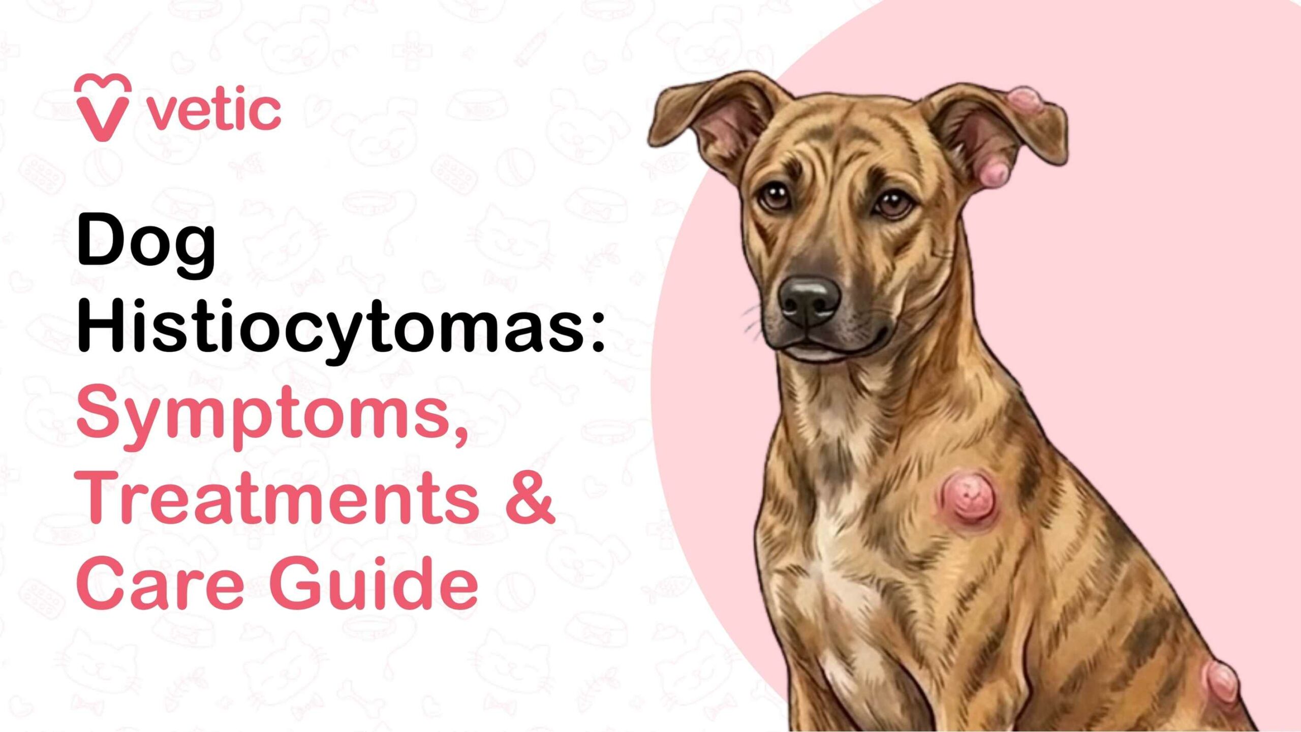
- A round, raised, button-like lump
- Smooth surface
- Pink to red in colour
- Firm but not usually painful
- Often appears suddenly and grows quickly over days to weeks
They are most commonly found on the head and ears, legs and paws, neck, and occasionally the trunk.
Because they are hairless and red, many pet parents initially mistake them for insect bites, warts, or even more serious tumours.
In some cases, the surface may ulcerate (break open), especially if the dog scratches or rubs it. Ulceration can make the lump look more concerning than it actually is.
Suggested Read: Scabies in Dogs: The Signs, Causes, and Treatment of Sarcoptic Mange (Mites) in Dogs
Common Symptoms of Histiocytomas in Dogs
In many cases, the only symptom is the visible lump itself. However, additional signs of histomas in dogs may include:
- Mild redness or swelling around the growth
- Occasional itching or licking
- Scabbing, if irritated
- Rare bleeding if traumatised
Most dogs do not show pain, lethargy, or systemic illness from a simple histiocytoma. If your dog seems unwell, another diagnosis should be considered.
In such cases, it is best to consult a vet online so your dog can be examined and appropriately investigated.
Causes of Histiocytomas in Dogs
The exact cause of histiocytomas in dogs is not fully understood. But several factors are believed to play a role:
- Immune system activity: These tumours originate from immune cells and often regress spontaneously. This happens because the dog’s immune system recognises the abnormal cells and attacks them, leading to tumour shrinkage and disappearance.
- Age-related immune response: Young dogs appear more susceptible, possibly due to an immature or highly reactive immune system.
- Genetic predisposition: Certain breeds are more prone to histiocytomas.
Importantly, histiocytomas are not caused by infection, injury, or poor hygiene. They are not contagious.
Suggested Read: Signs, Types, Diagnosis, Treatment and Outcome of Cancer in Dogs
Are Certain Dog Breeds More Prone to Histiocytomas?
Yes. While histiocytomas can occur in any dog, they are more commonly seen in certain breeds, including Boxers, Dachshunds, Cocker Spaniels, Labrador Retrievers, and Staffordshire terriers.
Young dogs of these breeds are especially likely to develop the classic solitary “button tumour.” However, breed predisposition does not mean a dog will definitely develop histiocytomas. But it can increase the likelihood.
How Veterinarians Diagnose Canine Histiocytoma
Even though histiocytomas often have a characteristic appearance, veterinarians do not rely solely on visual inspection. Many skin tumours can look similar, including malignant ones.
Diagnostic steps may include:
Physical Examination
Your veterinarian will evaluate:
- Size
- Location
- Texture
- Growth rate
- Whether the lump is fixed or movable
- Age of the dog
Fine Needle Aspiration (FNA)
This is the most common initial test. A small needle is inserted into the lump to collect cells, which are then examined under a microscope. This is often sufficient to diagnose a histiocytoma.
In many cases, FNAC can confirm a histiocytoma.
Biopsy
If the diagnosis is uncertain or the mass behaves atypically, a biopsy may be recommended. This involves surgically removing part or all of the mass and sending it to a pathology lab.
Biopsy is particularly important if:
- The dog is older
- The mass continues growing
- The mass looks aggressive
- The diagnosis is unclear
Treatment Options for Histiocytomas in Dogs
Canine histiocytomas treatment depends on:
- The dog’s age
- Tumour size and location
- Whether the mass is ulcerated
- Diagnostic confirmation
- Owner preference
In many cases, minimal intervention is needed.
Monitoring and Watchful Waiting
The most common and appropriate approach is monitoring. Why watchful waiting works:
- Many histiocytomas regress spontaneously within 2-3 months
- The immune system gradually eliminates the tumour
- Avoids unnecessary surgery in young dogs
During monitoring, vets recommend:
- Measuring the lump weekly
- Watching for ulceration or infection
- Preventing licking or chewing
If the mass shrinks over time, no further treatment for canine histiocytoma is usually needed.
Surgical Removal of Dog Histiocytomas
Surgery may be recommended if:
- The tumour does not regress after several months
- The mass is ulcerated, infected, and bleeding
- The dog persistently traumatises the area
- The diagnosis is uncertain
- The dog is older
Surgical removal is typically curative, with a low risk of recurrence when the mass is completely excised.
Medical and Topical Treatments
There is no specific medication that “cures” histiocytomas. But supportive treatments may be used:
- Anti-inflammatory medications
- Topical therapies to reduce irritation
- Antibiotics if secondary infection develops
- Bandaging in some cases
These treatments for dog histiocytoma in dogs do not remove the tumour. But it may help manage complications during regression.
In rare situations where the immune system does not trigger regression, additional therapies may be considered.
Suggested Read: Excess Foot Licking and Itching in Dogs: Pododermatitis
Home Care and Aftercare for Dogs With Histiocytomas
Proper home care supports healing and prevents complications:
- Prevent licking or chewing using an Elizabethan collar if needed
- Keep the area clean and dry
- Avoid applying human creams or home remedies
- Take weekly photos to track progress
- Monitor size, colour, and surface changes
- Follow up with your vet if the lump changes suddenly
After surgical removal, follow all post-operative instructions carefully:
- Prevent activity that may stress stitches
- Administer prescribed medications
- Watch for swelling, discharge, or redness
Most dogs recover quickly after surgical removal, especially if the tumour was small.
Recovery Timeline and Long-Term Outlook
The prognosis for dogs with histiocytomas is excellent.
Typical timelines:
- Rapid growth phase: 1-4 weeks
- Stable phase: 2-4 weeks
- Spontaneous regression: 4-12 weeks
- Post-surgical recovery: 10-14 days
- Long-term outcome: Full recovery with minimal risk
Once resolved, most dogs live normal lives without further issues related to that tumour.
Can Histiocytomas in Dogs Be Prevented?
There is no known way to prevent histiocytomas in dogs. This is because they are linked to immune and genetic factors rather than environmental or care factors.
What you can do:
- Regular grooming
- Perform regular skin checks
- Seek early veterinary evaluation for new lumps
- Avoid assuming all growths are benign
Early assessment prevents delays in diagnosing more serious conditions.
When a Histiocytoma May Be Something More Serious
Not every red, hairless lump is a histiocytoma. Some more serious tumours can look similar, especially in the early stages.
One important condition to rule out is a mast cell tumour. These tumours can vary widely in appearance and may resemble histiocytomas.
Warning signs that suggest a different or more serious tumour include:
- Mass changes size rapidly (shrinks and swells unpredictably)
- Multiple lumps appearing at once
- Severe swelling, pain, or discharge
- Lack of regression after 2-3 months
- Occurrence in older dogs
- Causes systemic signs (vomiting, lethargy)
In these cases, further testing is essential. Never assume a lump is harmless without a vet evaluation at the best pet clinic.
Final Takeaway: About Histiocytomas in Dogs
Histiocytomas in dogs are common, benign skin tumours, especially in young dogs. Although they can look alarming, most resolve on their own with time and monitoring. The key is not to self-diagnose. Every new lump should be checked by a vet to rule out more serious conditions. With proper evaluation and care, dogs with histiocytomas typically recover fully and comfortably.
FAQs: About Histiocytomas in Dogs
Is there a home treatment for histiocytomas in dogs?
No safe home treatment can remove histiocytomas. Because most regress naturally, “home treatment” usually involves careful monitoring rather than active intervention.
How quickly do histiocytomas grow in dogs?
Histiocytomas often grow rapidly over 1-4 weeks. This fast growth can be alarming but is typical.
Can histiocytomas in dogs come back after treatment?
Recurrence at the same site after complete surgical removal is rare. However, some dogs, especially predisposed breeds, may develop a new histiocytoma at a different location later in life.
How can you tell the difference between a mast cell tumour and a histiocytoma?
They can look similar. But, histiocytomas are more common in young dogs and often have a smooth, button-like appearance. Only cytology or biopsy can reliably distinguish between them.
Are histiocytomas painful for dogs?
Most histocytomas are not painful, though ulcerated or irritated lesions may cause discomfort. If your dog is licking, scratching, or showing signs of discomfort, consult your vet.
References
Bauhaus, J. (2025, November 3). Lumps and Bumps on Dog’s Skin: Signs, Symptoms, Causes. American Kennel Club. https://www.akc.org/expert-advice/health/dog-skin-lumps-bumps/
Brooks, W. (2017). Histiocytoma is a Benign Skin Growth in Dogs. VIN.com. https://veterinarypartner.vin.com/default.aspx?pid=19239&id=4952066
Brooks, W. (2026). Benign Sebaceous Gland Tumors. Vin.com. https://veterinarypartner.vin.com/default.aspx?pid=19239&catId=254091&id=4952215
Pires, I., Rodrigues, P., Alves, A., Silva, F., & Lopes, C. (2024). Histopathological and Ultrastructural Study of a Canine Langerhans Cell Tumour (Canine Cutaneous Histiocytoma). Cells, 13(15), 1263. https://www.mdpi.com/2073-4409/13/15/1263 Villalobos, A. E. (n.d.). Tumors of the Skin in Dogs. Veterinary Manual. https://www.msdvetmanual.com/dog-owners/skin-disorders-of-dogs/tumors-of-the-skin-in-dogs
