- Low albumin causes fluid to leak into the abdomen, chest, and limbs, visible as a swollen belly and oedema
- The most common causes are inflammatory bowel disease (IBD) and intestinal lymphangiectasia
- Diagnosis requires serum albumin testing, faecal protein loss testing, and intestinal biopsies
- PLE is managed rather than always cured; dietary control is central to every treatment plan
What Is Protein-Losing Enteropathy (PLE) in Dogs
Canine protein-losing enteropathy is a syndrome: a group of conditions sharing the same outcome, excessive protein loss through the intestinal wall.
Albumin, the main protein in blood, maintains oncotic pressure and helps keep fluid within blood vessels. When albumin drops too low, fluid leaks into the abdomen, the chest, and under the skin.
How the Digestive Tract Loses Protein
In a healthy dog, protein circulates in the blood and is only released into the intestine in small, controlled amounts. In protein-losing enteropathy in dogs, the intestinal wall becomes abnormally permeable. Albumin and other plasma proteins leak from the bloodstream into the intestinal lumen faster than the liver can replace them.
The result is hypoalbuminemia in dogs, a persistently low blood albumin level that drives all the clinical signs of PLE.
Common Causes of PLE in Dogs
Several intestinal diseases can cause excessive protein loss in dogs.
Inflammatory Bowel Disease (IBD)
IBD disease in dogs is the most common cause of PLE. Canine IBD is an immune-mediated condition that inflames the intestinal mucosa. Lymphoplasmacytic enteritis is the most frequently seen form. It damages the intestinal villi and increases gut wall permeability, allowing protein to leak into the intestine.
Predisposed breeds include Soft Coated Wheaten Terriers, German Shepherds, and Basenjis.
Intestinal Lymphangiectasia
Lymphangiectasia in dogs is the dilation of the lymphatic vessels within the intestinal wall. Lymph, which is rich in proteins and fats, leaks into the intestinal lumen instead of being absorbed normally. Yorkshire Terriers are among the breeds most commonly affected.
A strict low-fat diet is essential for managing lymphangiectasia. Dietary fat stimulates lymph flow and directly worsens protein loss.
Intestinal Tumours and Other Conditions
Intestinal lymphoma is the most common intestinal tumour in dogs and a significant cause of PLE. Other tumours and severe infections (e.g., parvovirus, hookworm) can also disrupt the gut wall, causing protein loss.
Symptoms of PLE in Dogs
PLE symptoms in dogs develop gradually but can become severe. Watch for:
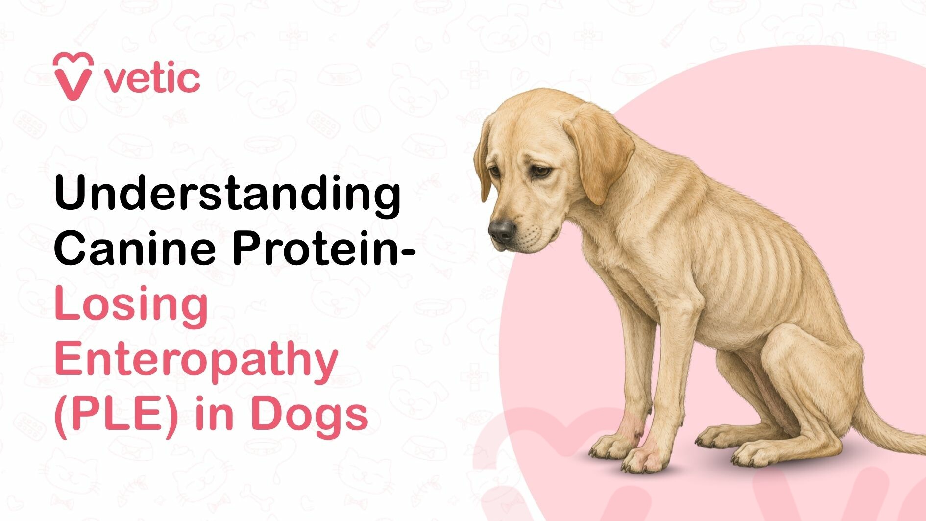
- Weight loss despite a normal or even increased appetite
- Chronic diarrhoea, which may be intermittent at first
- Vomiting
- A visibly distended or pot-bellied abdomen (ascites: fluid accumulating in the abdomen)
- Swelling of the face, legs, or limbs (subcutaneous oedema)
- Difficulty breathing (pleural effusion: fluid around the lungs)
- Lethargy, weakness, and exercise intolerance
- Poor coat condition
Why PLE Can Become a Serious Medical Condition
Hypoalbuminaemia in dogs disrupts far more than fluid balance. This type of protein enteropathy in dogs also causes:
- Hypocalcaemia: calcium is bound to albumin, so falling albumin pulls calcium levels down with it
- Coagulopathy: clotting factors are proteins that also leak through the damaged gut wall
- Pulmonary thromboembolism: Dogs with PLE face a high risk of life-threatening blood clots in the lungs, particularly Soft Coated Wheaten Terriers
- Progressive malnutrition and immune compromise in chronic, untreated cases
These complications are why PLE is a potentially life-threatening condition, not just a digestive problem.
How Vets Diagnose Protein-Losing Enteropathy
Diagnosis of PLE in dogs begins with blood tests. A low serum albumin (below 2.0 g/dL) combined with low total protein points to significant protein loss. The next step is determining whether the loss is coming from the gut or the kidneys.
Faecal alpha-1 protease inhibitor (fA1PI) testing specifically measures protein lost through the GI tract. Abdominal ultrasound assesses intestinal wall changes, lymph node size, and fluid accumulation.
Endoscopy with intestinal biopsies is the gold standard. Histopathology of the biopsy tissue identifies IBD, lymphangiectasia, or lymphoma.
Treatment Options for Canine PLE
Treatment of PLE in dogs depends on the underlying cause, but dietary change and medical management work together in almost every case.
Dietary Management
Diet is central to managing PLE disease in dogs regardless of the underlying cause.
- For lymphangiectasia, an ultra-low-fat diet (10-15% fat on a dry-matter basis) is non-negotiable. Dietary fat stimulates lymph flow and directly worsens protein loss.
- For IBD, a hydrolysed protein or novel protein diet reduces antigenic stimulation of the inflamed gut. Small, frequent meals put less demand on the damaged intestinal wall.
Medications and Supportive Care
IBD-related PLE is treated with immunosuppressives. Prednisolone is the first-line choice. Dogs that do not respond may be stepped up to azathioprine, chlorambucil, or cyclosporine.
Cobalamin (vitamin B12) supplementation is often needed because chronic enteropathy can deplete B12 stores. Plasma or albumin infusions may be required acutely when albumin is critically low. Dogs at high risk of thromboembolism may require preventive anticoagulation.
Intestinal lymphoma requires vet oncology consultation and chemotherapy.
Recovery, Monitoring, and Long-Term Outlook
Managing dog PLE requires long-term commitment from both the vet team and the pet parent. Albumin levels are checked every four to eight weeks initially to assess the response to treatment.
Diet typically needs to be maintained lifelong. Dogs with IBD-related PLE can achieve remission and live well for years with the right management. Lymphangiectasia requires permanent dietary control but is compatible with a good quality of life in many cases.
What Is the Life Expectancy of a Dog With PLE?
Life expectancy depends heavily on the underlying cause.
- IBD: With good dietary and medical management, many dogs live for several comfortable years after diagnosis.
- Lymphangiectasia: variable. Yorkshire Terriers and other susceptible breeds can often be managed effectively for years.
- Intestinal lymphoma: median survival is typically six to twelve months with treatment. This varies by lymphoma type and individual response to chemotherapy.
Early diagnosis and a clear management plan designed by a pet health expert give any dog with PLE the best possible outcome.
Final Thoughts: About Dogs PLE
PLE in dogs is manageable in many cases, but it demands a long-term veterinary partnership. No single medication or diet change is a quick fix. Consistent monitoring and a stable dietary routine make the biggest difference to outcomes.
If your dog is losing weight, developing a swollen belly, or has chronic unexplained diarrhoea, a Vetic vet can run the right tests and build a management plan. Book a consultation at your nearest pet clinic.
FAQs: About Protein-Losing Enteropathy in Dogs
Is PLE in dogs curable?
PLE in dogs is not always curable, but it is often manageable. When IBD is the underlying cause, remission is achievable with medication and dietary change. Lymphangiectasia requires permanent dietary management. Intestinal lymphoma is managed rather than cured.
What foods are recommended for dogs with PLE?
An ultra-low-fat diet is essential when the cause is lymphangiectasia. For IBD-related PLE, a hydrolysed protein or novel protein diet is recommended. Small, frequent meals reduce strain on the damaged gut.
Can dogs live a normal life with PLE?
Many PLE dogs live well with consistent management. Strict dietary control, regular monitoring, and appropriate medication allow most dogs to maintain a good quality of life.
Is PLE the same as IBD?
No. IBD is one of the most common causes of PLE, but PLE is a syndrome, not a specific disease. Intestinal lymphangiectasia, intestinal tumours, and severe infections can also cause PLE.
References
Collier, A. (2021, January). Chronic Enteropathies in Small Animals. MSD Veterinary Manual. https://www.msdvetmanual.com/digestive-system/diseases-of-the-small-intestine-in-small-animals/chronic-enteropathies-in-small-animals
Marks, S. L. (2026). Diagnosis and Management of Protein-Losing Enteropathies. VIN. https://www.vin.com/apputil/content/defaultadv1.aspx?pId=11242&id=3860867
Willard, M. (n.d.). Protein-Losing Enteropathy In Dogs And Cats. VIN. https://www.vin.com/apputil/content/defaultadv1.aspx?pId=11196&catId=30751&id=3854206